
Magnetic Particle Imaging
Nowadays there are several imaging techniques in clinical application. MRI and CT, for example, can both work without the need for an additional contrast agent. In comparison to that there are other imaging techniques such as PET or SPECT which only work by using a tracer. The advantage of imaging a tracer is that no tissue is visible, which leads to a high contrast.
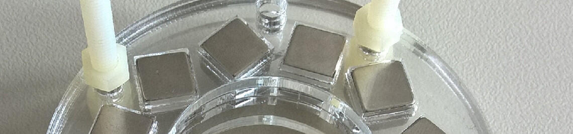
Since 2005 a new tracer method for imaging magnetic particles is known. It is called Magnetic Particle Imaging (MPI). The applied particles are superparamagnetic and composed of an iron oxide core with a biocompatible shell. They are called superparamagnetic iron oxides (SPIOs). They emit no harmful radiation for animals or humans in opposition to the nuclear tracers which are used in PET or SPECT. SPIOs are already used in MRI as contrast agents, which makes it easier to use them for the current research and future clinical applications. But there has to be put a lot of effort in creating the optimal particles for MPI.
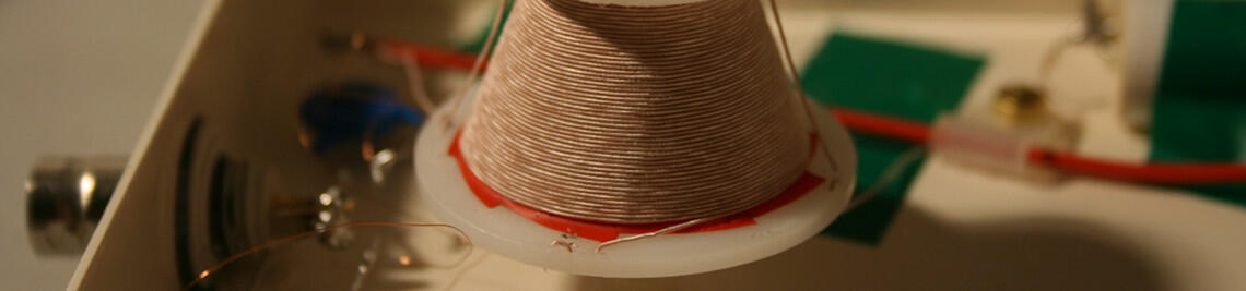
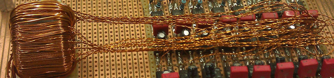
One can imagine the SPIOs as tiny permanent magnets. Applying an external magnetic field their orientation can be changed and the signal induced in a receiver coil by this change can be detected. This effect is used in Traveling Wave MPI. Another approach to manipulate these particles is to expose them to a rotating magnetic field and observe their relaxation behavior in Rotational Drift Spectroscopy.
Group leader: Prof. Dr. Volker C. Behr