Equipment
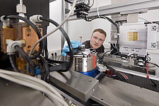
X-Ray Microscope XRM II
Source: Customized JEOL JSM-7100F Vacc 30kV Imax 400nA
Reflection Target: Nanostructured molybdenum and tungsten
Detector: Photon-counting direct-converting X-ray detector with 1024x512 pixels
Magnification: up to 1000 Resolution: down to 50nm Fully 3D imaging possible through piezo-powered rotational axis
Scanning Electron Microscope
SEI Resolution: 1.2 nm @ 30kV
Magnification: 10 to 1 000 000
Accelerating Voltage: 0.2 to 30 kV
Current: 10^-12 to 4*^10^-7 A
Electron Gun: In-lens Schottky field emission gun
Source: Rigaku microfocus-source with rotating anode (capacity: 1,2 kW)
Multilayer: Parabolic Goebel mirror for the Cu-Kα-line at 1.54 Å
Collimators: 3 motorised collimators each with 4 individually mobile hybrid-blades (monocrystal)
Axes: 3-linear axes each for detector- and positioning of sample with an additional 360°-rotation achse for rotatability of sampler
Sample Environment: Holding device for up to 8 solid samples, capillary holding device for up to 10 capillaries, holding device with heating for 1 capillary (temperature range: -5 – 80 °C)
Detectors: Dectris EIGER 1M (1035 x 1065 Pixel, Pixel size: 75 µm)
Liquid metal jet anode (LMJ)
The experiment is set up in an inter-locked x-ray hutch with 6 mm lead-shielding. The sample stage and the detector are built on a vibration damped granite base, both their axes are motorized. The maximum focus-detector distance equals 1.40 m.
Source: Excillum JXS D1 with specs:
- 70 kV max. acceleration voltage
- 200W max. output power
- 10 mu min. electron focus
- Target: liquid Ga-In-Sn alloy
Sample stage
- 2 circle goniometer (X-Huber, Germany)
- sample lift (X-Huber, Germany)
- sample translation (Micos PI GmbH, Germany)
- continuous rotation (Leuven Air Bearing, Belgium)
- piezoelectric 2-axis fine positioning (atocube, Germany)
Scintillator-based indirect detector
- P43 high-resolution phosphor screen (Proxivision, Germany)
- Rodenstock XR-Heliflex lens
- Nikkor Telephoto lens
- Andor NEO sCMOS detector (2180 x 2560 pixels, 100 fps)
- max. field of view 33.3 mm x 28.3 mm (13 mu pixelsize)
Tasks
- phase contrast and holo-CT of biological specimen
- in situ fast CT for materials testing / fatigue
- novel detector development (high-res and high-speed)

![[Translate to Englisch:] [Translate to Englisch:]](/fileadmin/_processed_/5/0/csm_Startseite_SAXSAnlage_V2_Fuchs_e4c1b4e130.jpg)

